

|
|
|
|
|
#1
 11.02.2010, 09:50:38
|
|||
|
|||
|
Citat:
 
__________________
„Ca o carpa lepadata toata dreptatea noastra” (Is 64,5) 
|
|
#3
11.02.2010, 10:10:38
|
|||
|
|||
|
Scanning electron micrograph of a human dendritic cell
 Scanning electron microscope image of a leaf from a Black Walnut tree. Image shows a cross-section of a cut leaf, itsupper epidermal layer, mesophyll layer with palisade cells and vascular bundles, and lower epidermal layer. The protrusion at center is just over 50 microns tall. (Dartmouth Electron Microscope Facility/Dartmouth College)# 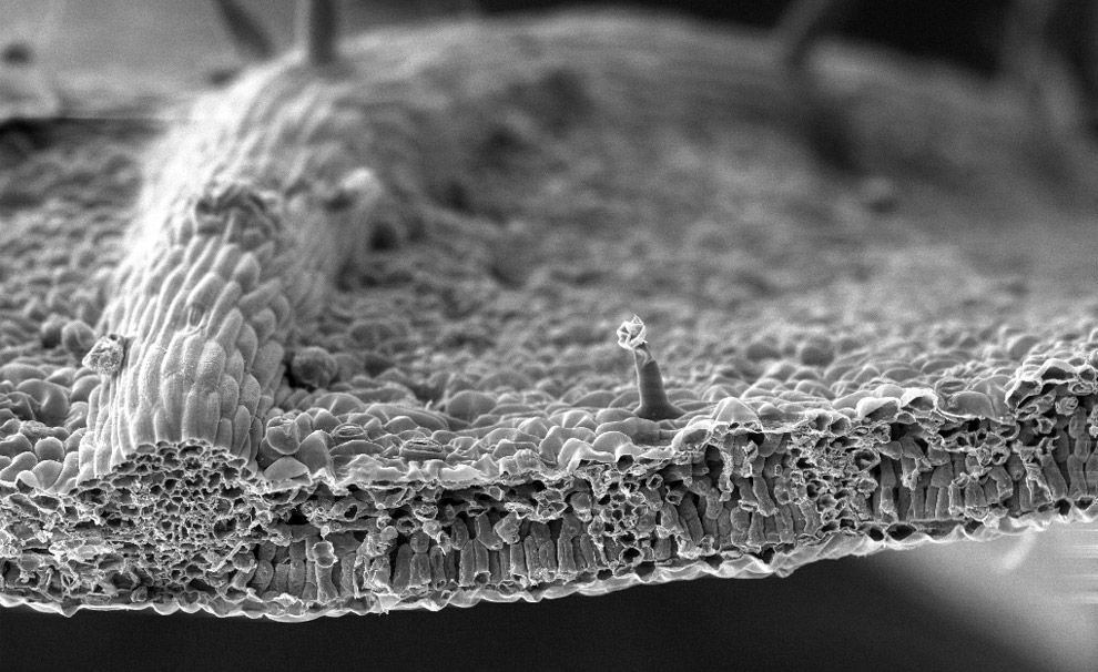 The anterior spiracles (respiratory openings) of a fruit fly larvae magnified 1500x. (Albert Tousson and Tomek Szul; Department of Cell Biology The University of Alabama at Birmingham)  A polllen grain on perched on the anther of a Penta lanceolata flower. The grain is about 40 microns wide. (Dartmouth Electron Microscope Facility/Dartmouth College) 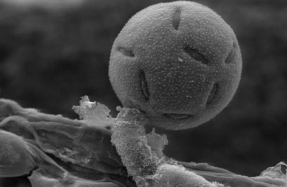 (Mai multe imagini aici: http://www.boston.com/bigpicture/200...cro_world.html )
__________________
„Ca o carpa lepadata toata dreptatea noastra” (Is 64,5)
|
|
#4
11.02.2010, 10:31:18
|
|||
|
|||
 Sperm production. Coloured scanning electron micrograph (SEM) of sperm cells (spermatozoa) in the seminiferous tubules of the testis. This is the site of spermatogenesis (sperm production). Each sperm cell consists of a head (green), which contains the genetic material that fertilises the female egg cell, and a tail (blue), which propels the sperm. The heads of the sperm are buried in Sertoli cells (yellow and orange), which nourish the developing sperm.  Title: SEM of human egg (ovum) surrounded by sperm in fallopian tube x550 Caption: Colour enhanced scanning electron micrograph
__________________
„Ca o carpa lepadata toata dreptatea noastra” (Is 64,5)
|
|
#5
11.02.2010, 10:35:41
|
|||
|
|||
 Coloured image of a 6 day old human embryo implanting (captured using a scanning electron microscope (SEM), a type of electron microscope that uses a beam of high-energy electrons to scan surfaces of images. The electron beam of the SEM interacts with atoms near or at the surface of the sample to be viewed, resulting in a very high-resolution, 3D-image)
__________________
„Ca o carpa lepadata toata dreptatea noastra” (Is 64,5)
|
|
#6
11.02.2010, 10:39:55
|
|||
|
|||
 (40 days)  (11 weeks) In 1969 Lennart Nilsson began using a scanning electron microscope to depict the body’s functions. He is generally credited with taking the first images of the Human Immunodeficiency Virus, and in 2003, he took the first image of the SARS virus.
__________________
„Ca o carpa lepadata toata dreptatea noastra” (Is 64,5)
|
|
#7
11.02.2010, 12:26:47
|
|||
|
|||
|
Citat:
Nu prea exista stiinta.Stiinta este un almagam de teorii care se bat cap in cap.Stiinta este in mare parte un rahat comestibil pentru prosti, care precum religia la randul ei se imparte in tabere, in care unii se bat cu altii si se contrazic cu altii.Stiinta contine prea putine adevaruri si enorm de multe teorii si supozitii prezentate ca fapte.
|
|
#8
11.02.2010, 15:42:40
|
|||
|
|||
|
Fotografiile sunt frumoase.
Dar majoritatea sunt doar interpretari computerizate ale unor date captate cu ajutorul telescoapelor si/sau radiotelescoapelor. Vreau sa spun ca norii aceia exista. Dar nimeni nu stie ce culori au. Culorile de redare sunt stabilite cu ajutorul calculatorului pe baza variatiilor de temperatura si a radiatiilor.
|
|
#9
11.02.2010, 15:46:50
|
|||
|
|||
|
Da, de-aia am pus "ochii" in ghilimele :)
Dar chiar si fara culori, detaliile sunt incredibile. Si pe mine ma frustreaza intr-o oarecare masura ca nu vedem lucrurile "asa cum sunt", dar ma consolez cu gandul "well, nu le vedem _acum_ si _aici_ :)
__________________
„Ca o carpa lepadata toata dreptatea noastra” (Is 64,5)
|
|
#10
11.02.2010, 15:51:23
|
|||
|
|||
|
Intradevar, pe masura ce avanseaza, stiinta ne dezvaluie imagini incredibile atat ale macrocosmosului cat si ale microcosmosului.
|
 |
|
 Subiecte asemănătoare
|
||||
| Subiect | Subiect început de | Forum | Răspunsuri | Ultimele Postari |
| "Prin complicităţi şi trădări, am ajuns să dăm judeţe întregi urmaşilor grofilor!" | Decebal | Stiri, actualitati, anunturi | 20 | 07.12.2013 19:56:55 |
| Ce vrea sa spuna acest citat prin "intrupam"? | cred_cu_adevarat | Generalitati | 2 | 13.10.2011 23:11:36 |
| Pelerinaje prin "Suflet pelerin" | laurschepsis | Pelerinaje la locuri Sfinte | 0 | 24.11.2010 08:31:11 |
| Care sunt "gaurile" stiintei? | Bast-et | Teologie si Stiinta | 28 | 19.09.2010 10:30:28 |
|
|


 Mod linear
