Scanning electron micrograph of a human dendritic cell

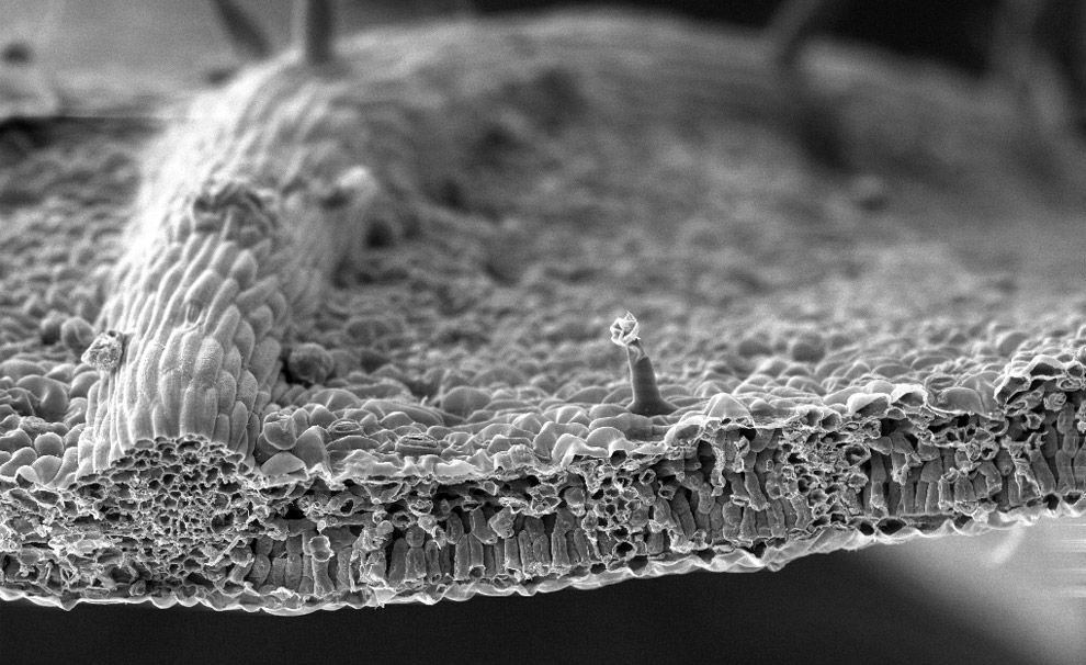
Scanning electron microscope image of a leaf from a Black Walnut tree. Image shows a cross-section of a cut leaf, itsupper epidermal layer, mesophyll layer with palisade cells and vascular bundles, and lower epidermal layer. The protrusion at center is just over 50 microns tall. (Dartmouth Electron Microscope Facility/Dartmouth College)#
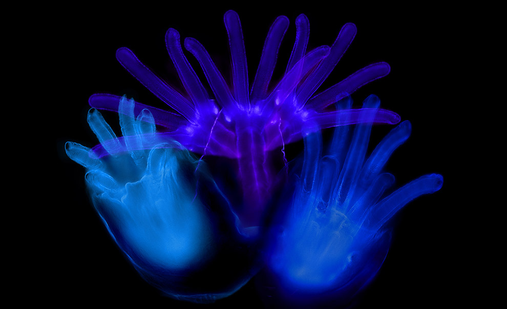
The anterior spiracles (respiratory openings) of a fruit fly larvae magnified 1500x. (Albert Tousson and Tomek Szul; Department of Cell Biology The University of Alabama at Birmingham)
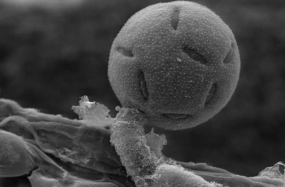
A polllen grain on perched on the anther of a Penta lanceolata flower. The grain is about 40 microns wide. (Dartmouth Electron Microscope Facility/Dartmouth College)
(Mai multe imagini aici:
http://www.boston.com/bigpicture/200...cro_world.html )